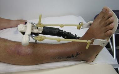

X-rays can reveal the presence of a fracture and the degree of displacement or fragmentation of the bone.ĬT scan: A CT scan is a more detailed imaging test that can provide a three-dimensional view of the bone and joint. X-rays: X-rays are typically the first imaging test performed to diagnose a Pilon fracture. They will also assess your range of motion, stability, and strength in the ankle joint. Physical examination: Your doctor will conduct a physical examination of your ankle to look for signs of swelling, bruising, and deformity. The following are the common methods of diagnosis for a Pilon fracture:
#Pilon fracture recovery blog skin
Open wound or visible bone protruding through the skin in severe cases.Swelling, bruising, or redness around the ankle.You should seek medical attention immediately if you experience any of the following symptoms: The longer you wait to seek treatment, the more difficult the fracture can be to treat, and the higher the risk of complications. If you suspect that you have a Pilon fracture, it is important to seek medical attention as soon as possible. Spiral fracture: Spiral fractures happen when the fracture spirals around the bone. Impacted fracture: An impacted fracture happens when the ends of the broken bone are driven into each other. Approximately 20% of pilon fractures are open fractures.Ĭomplete fracture: A complete fracture happens when the bone breaks into two pieces.ĭisplaced fractures: A displaced fracture means the broken bones do not stay aligned like they normally would be.Ĭomminuted fracture: A comminuted fracture means the bone breaks into more than two pieces. If the broken bone pierces through the skin, it’s called an open fracture or a compound fracture. Your healthcare provider may use one or more of the following terms to describe your pilon fracture:Ĭlosed or open (compound) fracture: If the fracture doesn’t break open the surrounding skin, it’s called a closed fracture.

There are also types of fractures that can apply to any bone break. Type C fractures are often associated with soft tissue injuries and can be very complex to treat. Type C: This is the most severe type of Pilon fracture and involves the fragmentation of the tibia bone and the ankle joint. Type B: This type of fracture is more severe and involves the fragmentation of the tibia bone into multiple pieces. Type A: This is the least severe type of Pilon fracture and involves a simple fracture of the tibia at the level of the ankle joint. There are different types of Pilon fractures, and they are classified based on the severity of the fracture and the degree of fragmentation of the tibia bone. This type of fracture involves a break in the tibia bone close to the ankle joint, which can cause significant pain, swelling, and bruising around the ankle. Once the cast or brace is removed, physical therapy is typically recommended to help restore strength, flexibility, and range of motion to the ankle joint.įrequently Asked Questions About Pilon Fractures Is a Pilon fracture painful?Ī Pilon fracture can be very painful.


The healing process can take several weeks or months depending on the severity of the fracture. After surgery, the ankle is immobilized in a cast or brace to allow the bones to heal. The surgeon may use screws, plates, or pins to hold the bone fragments in place while they heal. In most cases, surgery is required to realign the broken bones and stabilize the ankle joint. Treatment options may include surgery to realign and stabilize the broken bones, followed by a period of immobilization and rehabilitation to restore range of motion, strength, and function to the ankle. Pilon fractures are often complex injuries that require specialized treatment by an orthopedic surgeon.


 0 kommentar(er)
